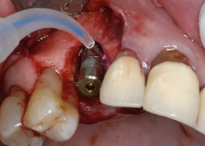
Stem Cells in Dentistry ?!?

Absolutely !!  Oral Surgery in general and bone grafting in particular are starting to make a transition.  More and more bone grafting scenarios employ cell-based or growth factor-enhanced grafting material.  Four items are absolutely essential in bone regeneration if predictable results are sought.

  • A matrix or scaffolding (collagen, bone mineral, synthetic grafts)
  • Cells (stem cells, platelets, osteoblasts)
  • Signaling molecules (growth factors, morphogens, adehsion molecules)
  • Time (often underestimated)

Within minutes after an “injury” to the bone structure, platelets aggregate in the area and release PDGF (Platelet Derived Growth Factor) and a variety of TGF-beta (Transforming Growth Factor – beta) molecules, to which BMPs (Bone Morphogenic Proteins) belong.  Some of the BMPs signal the Mesynchemal Stem Cells (MSCs) to “morph” into bone-precursor cells (osteoprogenitor cells).  Subsequently, PDGF signals these precursor cells to divide rapidly, in order to increase their number.  Once their number has increased (usually by an order of magnitude), a different set of BMPs will signal the precursor cells to “morph” again into mature bone-building cells (osteoblasts).

From this somewhat simplified molecular “injury cascade”, we can certainly appreciate the importance of stem cells.

A quick word on stem cells, because it has brought up some ethical and political issues in the past.  There are three types of stem cells in our body:

  • Embryonic Stem Cells (most potent, can form every tissue in our body)
  • Fetal Stem Cells (almost as potent, but somehwat more restricted in what they can become.  Often harvested from the umbilical cord and cryogenically frozen)
  • Adult Stem Cells (a.k.a. mesynchemal stem cell.  This cell is already committed to form only tissues of mesynchemal origin, i.e. bone, muscle, cartilage tissue, etc.).

The embryonic stem cells are the ones wich caused all the ethical controversy and to this day we can not perform any experiments with this cell lineage here in the U.S.  This cell line is extremely potent and can form any kind of tissue from all three primitive germ layers.

Our interest, however revolves around the Adult or Mesynchemal Stem Cells.  It stands to reason that an increased number of such stem cells during an “injury” or bone surgery can not only improve but also accelerate the bone healing.  It is now possible to use stem cell-fortified bone graft material (several thousand times the cell concentration of the human body), for various grafting procedures.  This graft material is very expensive and must be delivered within a day of surgery.  Initial results look very promising across several research studies.

Bookmark and Share


How many dental implants do I need to get rid of my denture?

This can be a tricky question.  First, I am going to assume that you want a fixed appliance or at least an appliance that gives you the same chewing power as a fixed appliance.  There are many people that do fine with removable dentures (plates), but then there are others, who really suffer with their dentures.  The empirical fact, however is this:  With a very well fitting pair of dentures (and that is very rare !!) a patient can achieve at most about 15% of their original biting power.  As you can imagine, this can severely limit your nutritional intake as well as your food enjoyment.  Dentures typically cover the whole palate.  Well, it just so happens that a great deal of taste buds are located in the palate, which are suddenly excluded from the tasting experience.

Now, in order to restore such a case to full functionality, we need several implants to gain enough bone anchorage in order to get the patient back to about 90% of their original biting force.  At a minimum 8 implants are recommended in the upper jaw and at a minimum 6 implants are recommended in the lower jaw (the bone is generally denser in the lower jaw !).

Additionally, the upper jaw may still need to be somewhat removable for the patient, so that he or she can clean the support structures for the appliance or prosthesis better.  This is not as often the case in the lower jaw.  The reason for this is that there is usually a flange on the lip side of the upper jaw prosthesis (see pictures below) and a little extension on the palatal side, to assure a good phonetic seal.  It is impossible to clean underneath this type of appliance if it was fixed.  We therefore often make “Fixed-Removable” appliances, which give you the same biting power as a completely fixed appliance, but can be removed for cleaning purposes.  Below are a series of images, which illustrate a “Fixed-Removable” appliance for the upper jaw.

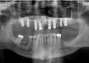
X-ray of 8 implants in the upper jaw

X-ray of 8 implants in the upper jaw

Here we see an x-ray image of implants placed into the upper jaw.  We chose 8 implants to give the patient enough support to be able to chew “steaks” again.  In this image one can see that the lower teeth need some extensive work too, but the patient wanted to get started with the upper arch first, because he could not tolerate his denture anymore.

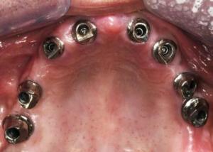
8 implants in the upper jaw with abutments connected

8 implants in the upper jaw with abutments connected

This image shows the implants in the patient’s mouth.  actually what is visible are the abutments, which are connected to the implants.  The implants themselves are buried in the bone and underneath the gum tissues and are therefore not visible.  These abutments, however are custom milled abutments which exhibit a 3-degree taper.  This is necessary, because the appliance will attach to these abutments via simple friction fit.
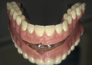
Fixed-Removable Appliance

Fixed-Removable Appliance

In this image we can see the appliance itself.  As you can see, it has only minimal extensions of acrylic and no palatal coverage.  The amount of pink acrylic necessary will largely depend on the amount of bone and gum tissue loss that has happened prior to the implant placements.  Once teeth are extracted, bone and gum tissue will shrink away.  That is an inevitable fact.  The less tissue loss we have, the less pink acrylic we have to use.
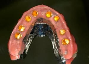
Palatal side of the Fixed-Retrievable appliance

Palatal side of the Fixed-Retrievable appliance

This image shows the flip side ( the palatal side) of the “fixed-retrievable appliance.  You can clearly see that it is metal re-enforced, in order to give it good stability and fracture resistance.  Furthermore, you can appreciate the “golden” metal caps.  These are the “female” friction components to the custom abutments shown above.  The patient will be able to positively seat the appliance without any rocking and will also be able to “wiggle” it back out.  The chewing power the patient gains is similar to that of a completely fixed bridge.  Additionally, the bone and gum tissue resorption has been arrested.  Since the dental implants are bone anchored devices, they transmit enough of a “stimulus to the bone, to maintain itself rather than resorb.
Please keep in mind that this represents only one out of many appliance options that could have been used in this scenario.  The one which is depicted here is a result of what worked best for this particular patient along several parameters.


Bookmark and Share


Can I get a dental implant if I take Coumadin?

Coumadin is an anticoagulant (blood thinner), which reduces the formation of blood clots. It does so by blocking the synthesis of certain clotting factors. A reduction in clotting factors will also reduce the chance of any blood clot formation.

Coumadin is predominantly used to prevent heart attacks, strokes and blood clots in veins and arteries as well as around prosthetic devices, such as artificial heart valves. The down side of Coumadin are the prolonged bleeding times. This is of great concern to anyone who needs to undergo minor surgery.
Many patients who take Coumadin get their INR (International Normalized Ratio) and PT (Prothrombin Time) tested on a regular basis. The Prothrombin time (PT) evaluates the ability of blood to clot properly, whereas the International Normalized Ratio (INR) is used to monitor the effectiveness of blood thinning drugs such as Coumadin (or also Jantoven, Marevan and Waran, which are all brand names for the generic Warfarin).

Most dental surgeons will look for the INR assessed a day prior to surgery, to determine whether it is safe to perform any minor oral surgical procedure, however some also consider the PT time along with the INR. Your cardiologist is always the final decision maker, however. Sometimes they will take you off the Coumadin and switch you over to Heparin a few days prior and after the surgery, sometimes they may just take you off for a couple of days. This will really depend on how high your risk of clot formation is and only your cardiologist can make this decision.

So to answer the original title question: As long as you are carefully monitored and prepared by your cardiologist just prior and after the dental implant surgery, you can get dental implants. There are no published studies showing an decrease in success rates of dental implants in patients taking any of the Warfarins.

Bookmark and Share


How Cold Lasers Work…

“Cold Lasers” seem to be the latest Buzz Word in Laser Dentistry. Cold Lasers are also known as “Soft Lasers” or, more scientifically, Biostimulatory Lasers.  As already mentioned in previous posts, these lasers do not cut or vaporise any tissue.  Although often very powerful, these lasers are defocused enough so that the tissue interaction is that of biostimulation and not thermal.

As we have seen earlier, cold lasers can be used for a variety of applications not only in dentistry, but also in medicine.  Their typical application field is that of tissue healing (Well, lately the application of hair re-growth has also been added and actually FDA approved, but there is little if any scientific substantiation to this at this point).

In order for laser light to be absorbed, there must be receptors.  Such receptors are well known in plants, but there are human light receptors other than those in the eyes and the skin.  In fact, to date more than three hundred photochemically reactive proteins, capable of harvesting low light energy, have been identified in both prokaryotic and eukaryotic organisms.  In humans, the most commonly known photochemically active receptor proteins are the rod and cone pigments in the eye.  However, other human photoreceptors have been discovered in recent times.  In fact today, we know that the majority of our cells have photoreactive molecules in them and we call those CHROMOPHORES.

It has been observed that if laser light is administered in the right dose, certain cell functions are being stimulated, and this is particularly evident if the cell in question has an impaired function.  It is known that laser light will cause certain chromophores in our cells to allow the build-up of radical oxygen species (singlet oxygen, instead of O2), which in turn influences the the formation of ATP (Adenosine Tri-Phosphate), which is the cells basic energy and fuel molecule.  Now, if the production of ATP reaches a certain level within cells, this will lead to a host of secondary effects, which have been studied and measured in several contexts:

  • increased cell metabolism and collagen synthesis in fibroblasts
  • increased DNA and RNA formation in the cell nucleus
  • increased cell division cycles
  • increased cell differentiation cycles of primitive cells (stem-type cells)

All of the above cellular effects will translate into faster healing by virtue of an increased population of cells which are involved in the inflammatory and healing cascades of the body.  Bone and soft tissue tend to heal faster and better after surgery, nerve cells can regenerate themselves at higher rates and pain is often reduced due to a laser-induced block of the pain receptor cells.

Bookmark and Share


Bone resorption of the Jaw

Continue reading

A 100 Watt Laser – And My Doctor Says It Won’t Hurt?!?!

It is an unfortunate business practice, but the sale of medical lasers is largely being promoted by their hardware profile: “X” number of watts, “Y” number of pulses at “Z” nanometers etc.  What is unfortunate about this is that it does not really paint a good picture on what the clinical qualities of a laser really are, because this type of advertising does not really address what kind of “tissue interaction” it produces.  This however is ultimately the most important quality of any laser.  I’ll explain…

First and foremost, the power of the laser (usually displayed in Watts) is the true output power in terms of light energy emitted.  This is in direct contrast to a light bulb for instance.  A 60-Watt light bulb will draw 60 Watts of power out of the socket, but only deliver a fraction thereof as light energy, because most of the power drawn gets converted to heat energy.  In a laser the power rating is NOT what it draws out of the electric socket, but rather the light energy it produces.

Another concept that needs to be addressed is that this power claim describing a laser, can often be misleading.  More often than not, a “high-powered” healthcare laser in the 20 – 100 Watt range achieves this kind or output power mostly in a “pulsed” mode.  This means that the laser will be “on” and “off” several hundred or even several thousand times a second and every time it is “on” it emits 100 Watts.  Since this is a pulse train of laser light, it is important to note that the AVERAGE power may only be in the milliwatt range, so there is effectively only less than 1 Watt being absorbed by the tissues.

The last and most important concept which needs to be addressed is that of the power density at the output tip (aka “fluence”).  A 50-Watt laser with an output diameter of 1 cm will have an entirely different effect on tissues than a 6-Watt laser with an output diameter of only a few hundred microns.  The former will have a biostimulatory effect, whereas the latter will be able to cut tissue.

So, as we can see, the advertising profiles of lasers do not really always reflect the clinical relevance.  It is my opinion that this needs to change eventually, so that the tissue interaction is placed into the foreground and not the hardware profile.


Bookmark and Share


Sinus Augmentation or Sinus Lift Procedure

The Sinus Augmentation of Sinus Lift Procedures are very common bone grafting procedures within the realm of implant dentistry.  Patients are often not quite clear what these procedures actually are, so I would like to

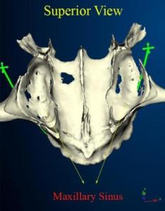
The maxillary sinus on a CT scan 3-D image

The maxillary sinus on a CT scan 3-D image

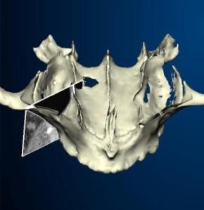
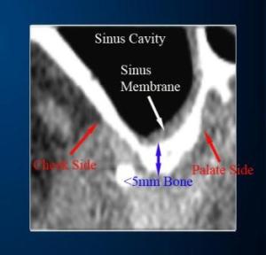
shed some light on these grafting procedures.  The maxillary sinus is the largest of all sinuses in the head-and-neck area.  It is located just to the inside of the bone in the upper cheek area.  The maxillary sinus starts out as a small air cavity in children and then expands and gets bigger as we grow older.  Once teeth are missing in the back areas of the upper jaw, the sinus will expand even further down towards the jaw ridge from the inside of the jaw bone.  This can be seen on the images on the right, especially the very bottom image, which shows a cross-sectional CT scan slice through the alveolar process of the upper jaw bone.

Now, as the sinus expands, and encroaches upon the alveolar ridge from the inside, you can appreciate that there is a diminishing distance of bone left between the top of the ridge and the floor of the sinus.  If this distance is too small to place one or several dental implants of proper length, we need to perform a sinus lift or sinus augmentation procedure, in order to re-gain this distance.

What is the difference between a “Sinus Augmentation” and a “Sinus Lift Procedure”?  Well, many will use these two terms

A CT Scan Slice through the sinus and alveolar process

A CT Scan Slice through the sinus and alveolar process

interchangeably, however there is a little difference between these two terms.  A “Sinus Augmentation” is a slightly more aggressive procedure, where a window is cut into the bony cheek side wall of the sinus and the sinus membrane is then gently lifted off the sinus floor, until a bone graft is finally placed underneath the lifted membrane.

A Sinus Lift procedure, on the other hand is usually performed right through the hole which is drilled for the implant(s).  No window is cut on the cheek side of the bone.  The Augmentation is usually done, for bigger lifts and the Lift procedure is usually done for smaller lifts.

So when the sinus membrane is finally lifted in either procedure, a bone graft is placed underneath the membrane, which keeps the membrane “tented” up.  For more detailed information on bone grafts see my bone graft post or link to Robert Gougaloff ‘s website.  This bone graft will then “mature” over the next six to 24 months (depending on the type of graft used).  What this

A CT Scan Cross sectional slice from the above image

A CT Scan Cross sectional slice from the above image

has accomplished however, is that the floor of the sinus was effectively “raised” and has thus given us enough room to place one or more dental implants of proper length.

Sinus Augmentations and dental implants can be done in one stage or in two stages, depending on how much residual alveolar bone was left to stabilize the implant.  If it is done in two stages, then the dental implant is usually placed 6 to 12 months following the sinus augmentation, depending on the graft material used.

Sinus Lift Procedures and the placement of dental implants are usually done at the same time, which obviously shortens the treatment time dramatically.  However this can usually be only done if the amount of lift needed is not too excessive.

A video of a Sinus Augmentation procedure can be seen on the following video link: Robert Gougaloff ‘s Sinus Augmentation Video.

Bookmark and Share


Osteoporosis, Bisphosphonates and Dental Implants

I have heard a lot of questions asked by dental professionals as well as patients regarding these topics lately.  The underlying question usually was: Do they mix?

Osteoporosis is a common disease characterized by decreased bone mass, increased bone turnover, and increased susceptibility to fracture.  In general, Osteoporosis itself is NOT a contraindication to having dental implants done.  Studies have shown that patients with osteoporosis enjoy the same kind of success rates with dental implants as patients without osteoporosis do.  What CAN present itself as a potential risk factor to dental implant surgery (and any kind of oral surgery for that matter), however, is the administration of Bisphosphonates (such as FOSAMAX®), which are designed to counteract the effects of osteoporosis.

Bisphosphonates belong to a class of drugs which inhibit osteoclast action and thus the resorption of bone.  This works well for maintaining good bone density (since the resorption cycle is being interrupted), however it can have disastrous consequences following bone surgery.  Bone will undergo a very structured healing response after surgery, which includes remodelling and turnover.  Osteoclasts play a very important part in this remodelling cycle of the healing phase.  If the osteoclast action is interrupted, osteonecrosis (bone cell death) may be a consequence.

Now not every regiment of bisphosphonates is a significant risk factor for post surgical osteonecrosis.  Up until very recently we have only haphazardly determined that injectable regiments of bisphosphonates or regiments of oral bisphosphonates for over three years present a definite contraindication to dental implant surgery.  Recently however, a test became available, which gives us a much more accurate way of determining the risk factor:  The CTx Test.

The CTx test, also known as the serum C-terminal telopeptide test, is a medical blood test that is used to assess the risk of bisphosphonate-induced osteonecrosis of the jaws.  C-terminal telopeptide is a marker used to measure bone metabolism. It is a by-product of normal bone metabolism or bone turnover.  If the CTx test shows a low value of CTx, then the implication could be that the bone turnover is low, thus the bone is less likely to recover from trauma, such as a tooth extraction or implant placement.  According to Marx ( J Oral Maxillofac Surg. 2007 Dec;65(12):2397-410.): “A stratification of relative risk was seen as CTX values less than 100 pg/mL representing high risk, CTX values between 100 pg/mL and 150 pg/mL representing moderate risk, and CTX values above 150 pg/mL representing minimal risk. The CTX values were noted to increase between 25.9 pg/mL to 26.4 pg/mL for each month of a drug holiday indicating a recovery of bone remodeling and a guideline as to when oral surgical procedures can be accomplished with the least risk.”   The latter portion of Marx’s statement implicates that any kind of Oral Surgery (including dental implant placement) could be an option, if a patient is taken off the regiment (under careful observation of his or her physician, of course) for about 3 months and a CTx re-test is taken for verification of new values.

If necessary, your dental professional or physician can order this test for you.  This test should be taken after 12 hours of fasting.

Bookmark and Share

A little bit on bone grafting…

The topic of bone grafting seems to be one of the more confusing areas in implant dentistry.  When do I need it, why do I need it and why is it so darn expensive?  Generally a bone grafting procedure is needed whenever there is insufficient bone available to place a dental implant of proper dimension.  Bone will resorb over time in the areas where teeth are missing.  I still don’t believe that patients always get the best explanation as to what the graft options are and what the pros and cons are – well, hopefully this little blog will shed some light on this topic.

For starters, let’s just quickly review the different classifications of bone grafts.  All bone graft materials can essentially be classified into the following classes:

  1. Autografts
  2. Allografts
  3. Alloplasts
  4. Xenografts
  5. Growth Factor Enhanced Grafts

Now, all of these five classes can have either or both of the following properties:

  • Osteoconductive – the ability to “guide” bone cells
  • Osteoinductive – the ability to accelerate bone regeneration

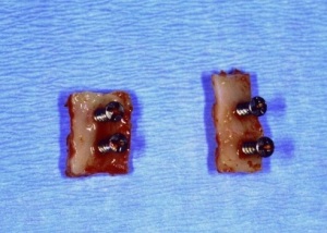
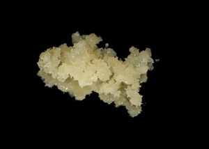
    A typical autograft from the chin

    A typical autograft from the chin

Autografts are bone grafts, which come from your own body.  This type of graft is still considered the “gold standard” by many surgeons.  It offers the best of both worlds; it has very good osteoconductivity and also great osteoinductivity due to its high content of resident growth factors.  The disadvantage is the higher morbidity.  It is always necessary to conduct a secondary surgical access in a remote location of the body in order to harvest the bone.  Depending on the quantity of bone needed, this can be the hip (for larger quantities) all the way down to an intra-oral site, such as the chin or the back of the jaw.  Autogenous bone grafts have shown to be some of the most predictable grafts in surgery.

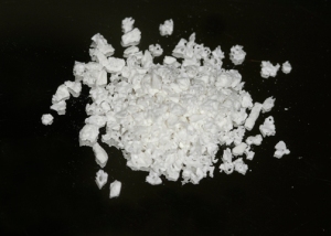
A typical allograft from a bone bank

A typical allograft from a bone bank

Allografts are a close relative of the autograft, in that it is of human origin, usually cadaver bone from a bone bank.  This always sets off a red flag with people with respect to disease transmission, however with the tight screening protocol and advanced processing technology this is virtually unheard of in the United States.  Allografts have the big advantage that they do not require a secondary surgical access site, however, they also have predominantly osteoconductive properties and very little if any osteoinductive properties.  Graft assimilation and maturation takes therefore longer than with the autografts.  Grafting success rates have also favored the autografts by a slight margin, depending on the individual application.

A typical xenograft of bovine origin

A typical xenograft of bovine origin

Xenografts are bone graft substitute from a different species all together, usually from bovine origin.  These grafts have usually only osteoconductive properties, since the organic portion has been completely removed (so no, there is no chance to contract BSE a.k.a. Mad Cow Disease).  The advantage of a xenograft is that there is a large quantity of bone available for the screening process for an exact micro architecture that is needed.  Their osteoconductive properties are therefore very good.  It can be distributed in particular form or as blocks.  Xenografts have shown to generate astonishing results, especially in sinus lift surgeries, which are often necessary for the placement of dental implants into the back areas of the upper jaw.

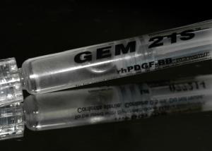
An example of recombinant human growth factor

An example of recombinant human growth factor

Growth Factor Enhanced Grafts are very new on the market.  These grafts capitalize on the regenerative powers of human growth factors such as Platelet Derived Growth Factors or Bone Morphogenic Proteins, both of which are generated by our own body during a bone repair and remodeling cycle, however these grafts are manufactured with recombinant DNA technology and are very concentrated.  These growth factor formulations are usually in liquid form that need to be combined with a “carrier”, such as collagen or a calcified matrix.  These grafts offer very good success rates, but their caveat is that they are VERY expensive.  This is an important point to consider, because more often than not, they just accelerate the graft assimilation and maturation time.  Research has yet to show whether the bone quality differs between all these different graft materials after a year or two.

Well, I hope this was informative for you.  I will be available for questions and comments, so don’t hesitate to post.

Bookmark and Share

So what can surgical lasers be used for in dentistry?

Surgical lasers are currently undergoing a tremendous evolution in dentistry.  Much of it has to do with the ability of manufacturers to scale their size down dramatically, and to make them more user friendly.  Additionally, new wavelength are constantly being added to the repertoire, which in turn broadens the

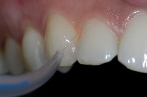
A periodontal procedure performed by a laser

A periodontal procedure performed by a laser

application field.  Dental or surgical lasers are defined by their lasing medium (the substance inside the laser, which creates the laser light), which in turn defines the wavelength of the emitted laser beam.

The following are the most common lasers used in dentistry:

  • Nd:YAG laser
  • Diode laser
  • ER:YAG laser & Er,Cr:YSGG laser
  • Carbon Dioxide laser

Each laser has its own unique application, based on the type of tissue interaction it offers.  Some lasers have their greatest energy absorption in soft tissue, others in bone tissue and yet others in hemoglobin or melanin.  the applications for lasers is basically chosen by their absorption spectrum in human tissue.

One of the main applications today in dentistry is soft tissue surgery.  Lasers are often used in periodontal disease cases, to complement traditional scaling and rootplaning procedures.  Lasers do not cause the kind of discomfort that traditional periodontal procedures cause.  That being said, not all periodontal procedures can be replaced by lasers either.

Another application which is gaining ground is photosterilization of compromised teeth or dental implants.

Photosterilization of a compromized dental implant

Photosterilization of a compromised dental implant

Often times we try to save compromised teeth and dental implants with a grafting procedure, however, a pre-requisite is that the surface of such a tooth or implant is meticulously cleaned and sterilized, or new bone will not form against it.

Laser bone surgery is just in its infancy in dentistry, but new powerful lasers make this possible already.  The advantage of laser bone surgery (in contrast to rotary instruments or piezo-electric instruments) is the cleaner and more sterile surgical field as well as the reduced post-operative inflammation and discomfort.

Lastly there is the new field of biostimualtion, which has actually nothing to do with surgery, but can help dramatically in the healing process.  Biostimualtion was already discussed in a previous post underCold Lasers.

For more information on lasers in dentistry, please visit my website at: http://www.robertgougaloff.com.